
Kidney Diagram Unlabeled Human Body Anatomy
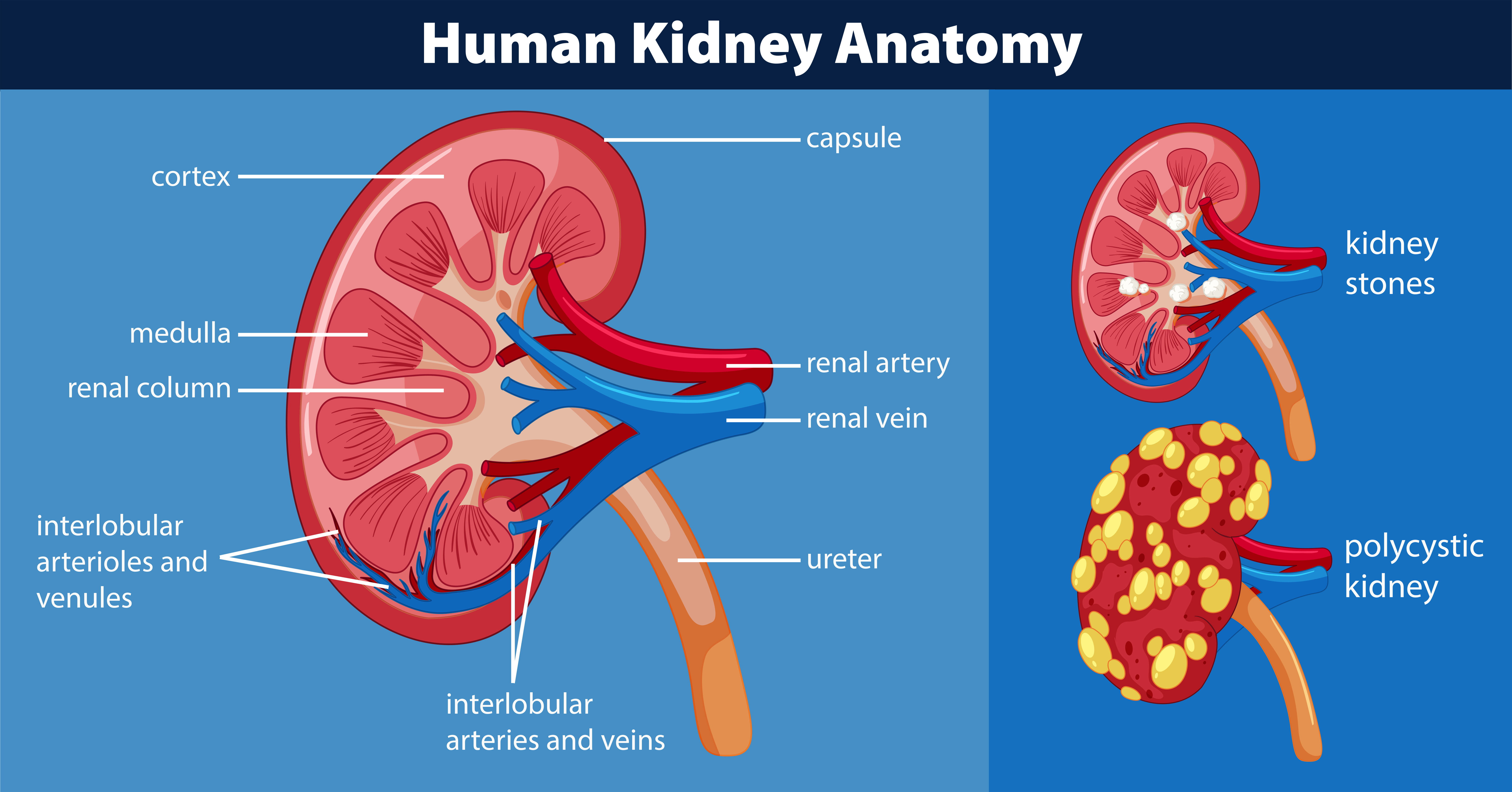
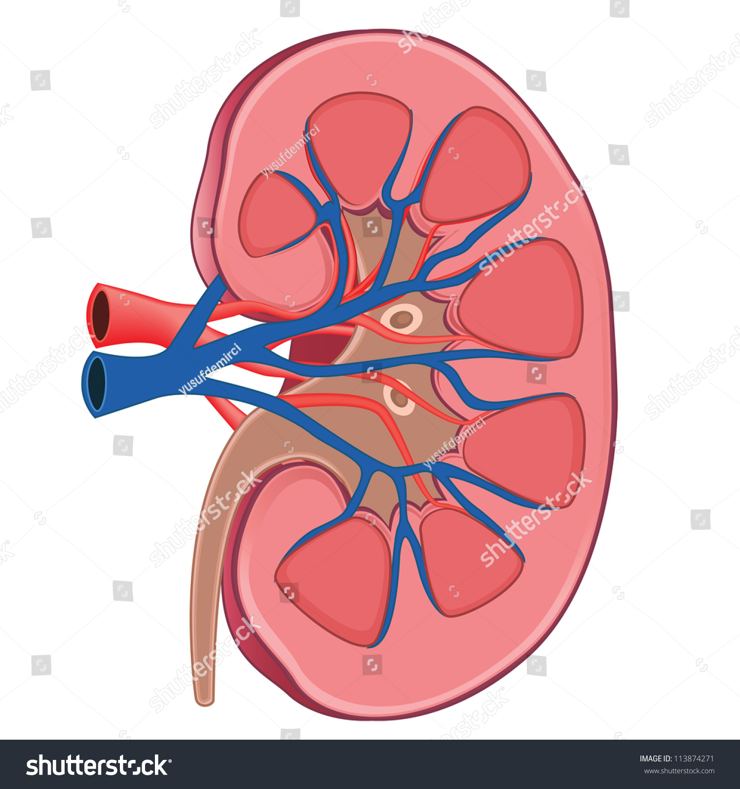
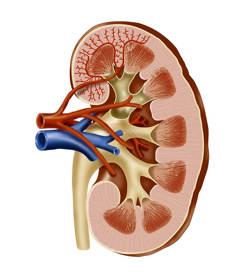
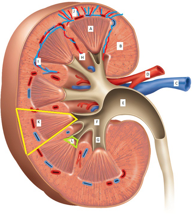
Kidney Anatomy. The shape of each kidney gives it a convex side and a concave side. You can see this clearly in the detailed diagram of kidney anatomy shown in Figure 19.3.3 19.3. 3. The concave side is where the renal artery enters the kidney and the renal vein and ureter leave the kidney.
Human kidney anatomy diagram 446409 Vector Art at Vecteezy
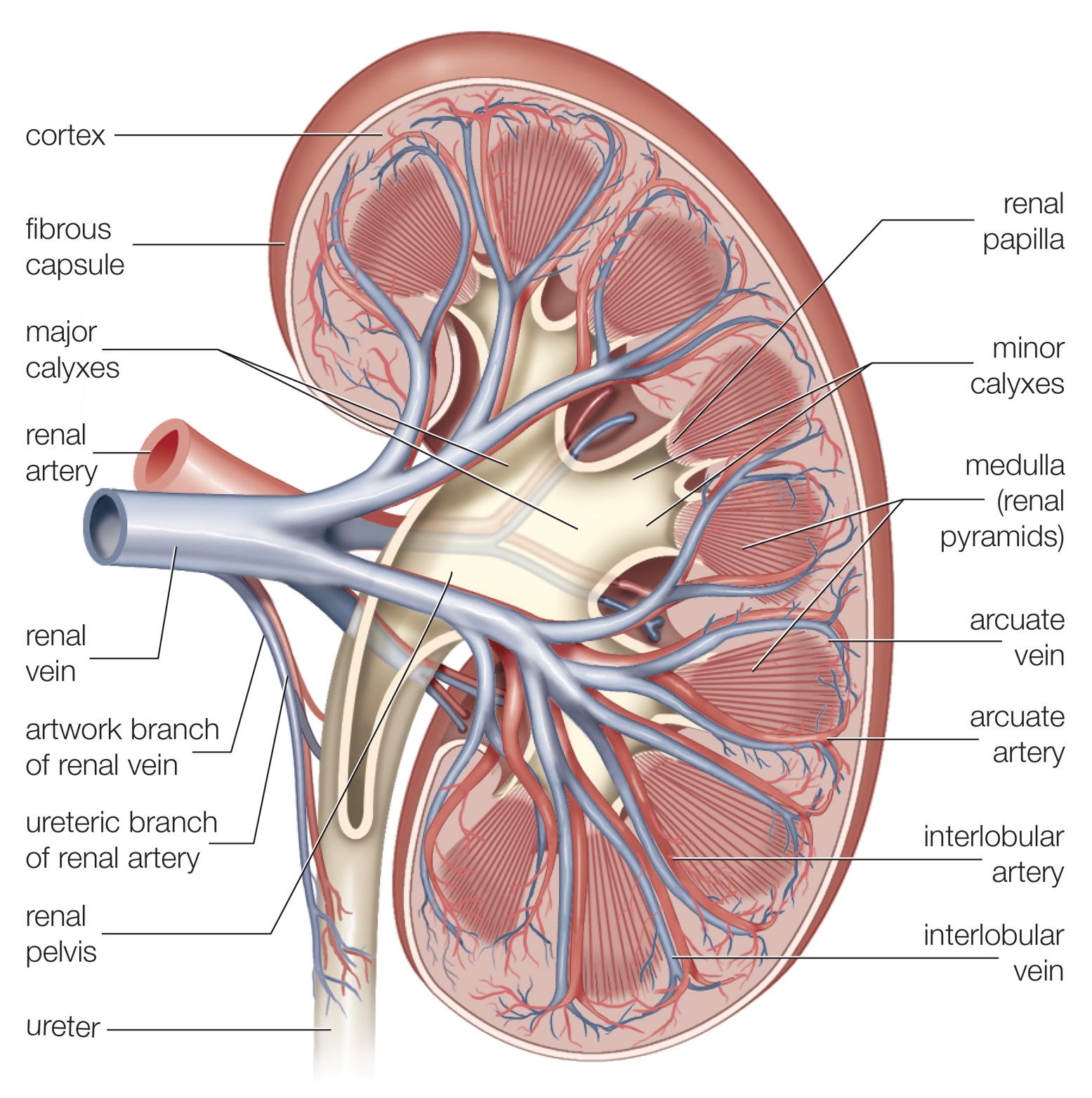
Kidney: Gross Anatomy (Illustrations Collection) MEDIA MENU Background Info A frontal section through the kidney reveals an outer, lighter-colored region called the renal cortex and an inner darker-colored region called the medulla. READ MORE Frontal Section DOWNLOAD IMAGE Unlabeled Version (1125px X 1150px) Terms of Use Renal Hilum DOWNLOAD SET

37 Color And Label The Urinary System Labels 2021
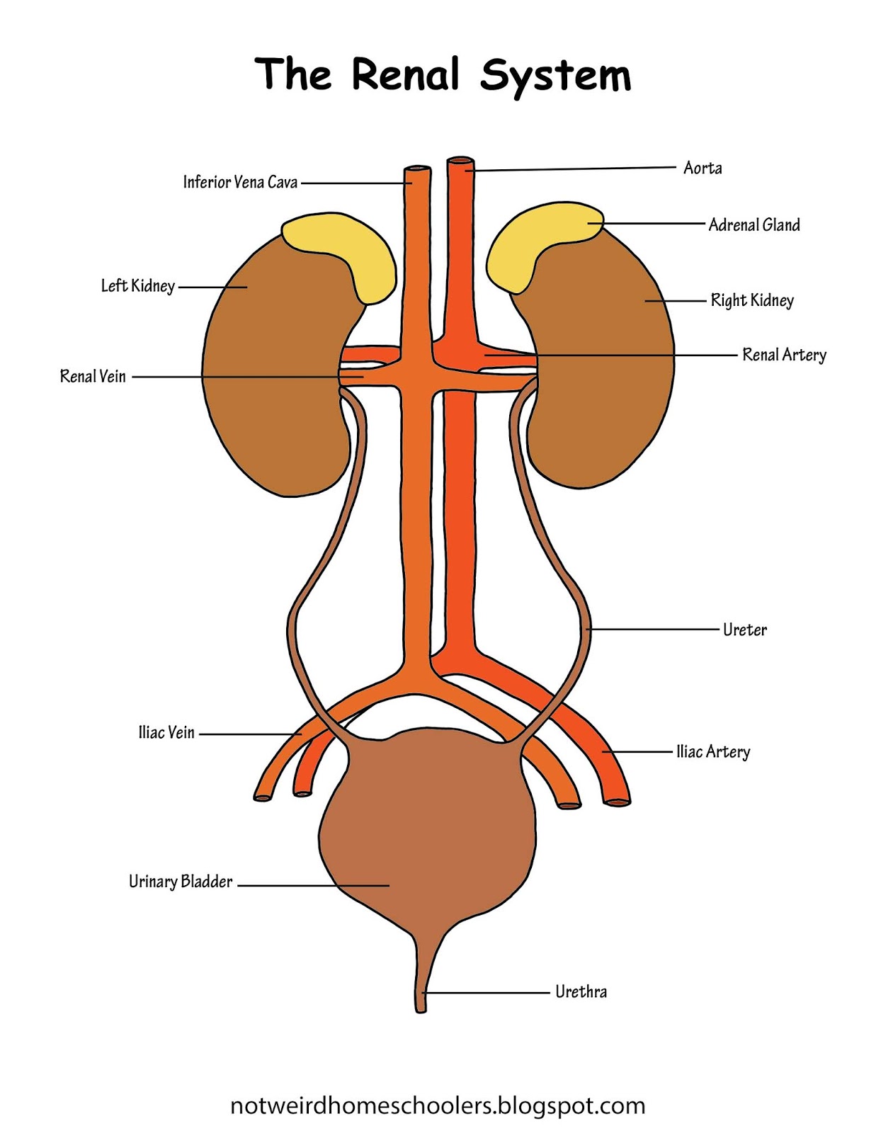
The urinary system is composed of a pair of kidneys, a pair of ureters, a bladder, and a urethra. These components together carry out the urinary system's function of regulating the volume and composition of body fluids, removing waste products from the blood, and expelling the waste and excess water from the body in the form of urine.
Kidney Diagram Unlabeled Human Body Anatomy
Anatomy of the Kidney Variant Image ID: 3512 Add to Lightbox. Save to Lightbox. Email this page; Link this page ; Print; Please describe! how you will use this image and then you will be able to add this image to your shopping basket. Pricing. Price for Add To Cart . 0 items.
What Are the Parts of the Human Kidney? Healthfully
Label and Color a Diagram of the Kidney Using Listed Terms Label and Color the Kidney This worksheet has a very simplified view of a kidney showing the cortex, renal pyramids, renal artery and vein, renal pelvis, and ureter. Students can practice labeling the structures and color coding the diagram.

Internal anatomy of the kidney Diagram Quizlet
The urinary system consists of the kidneys, ureters, urinary bladder, and urethra. The kidneys filter the blood to remove wastes and produce urine. The ureters, urinary bladder, and urethra together form the urinary tract, which acts as a plumbing system to drain urine from the kidneys, store it, and then release it during urination.
Medical and Health Science Kidney Internal Structure!!
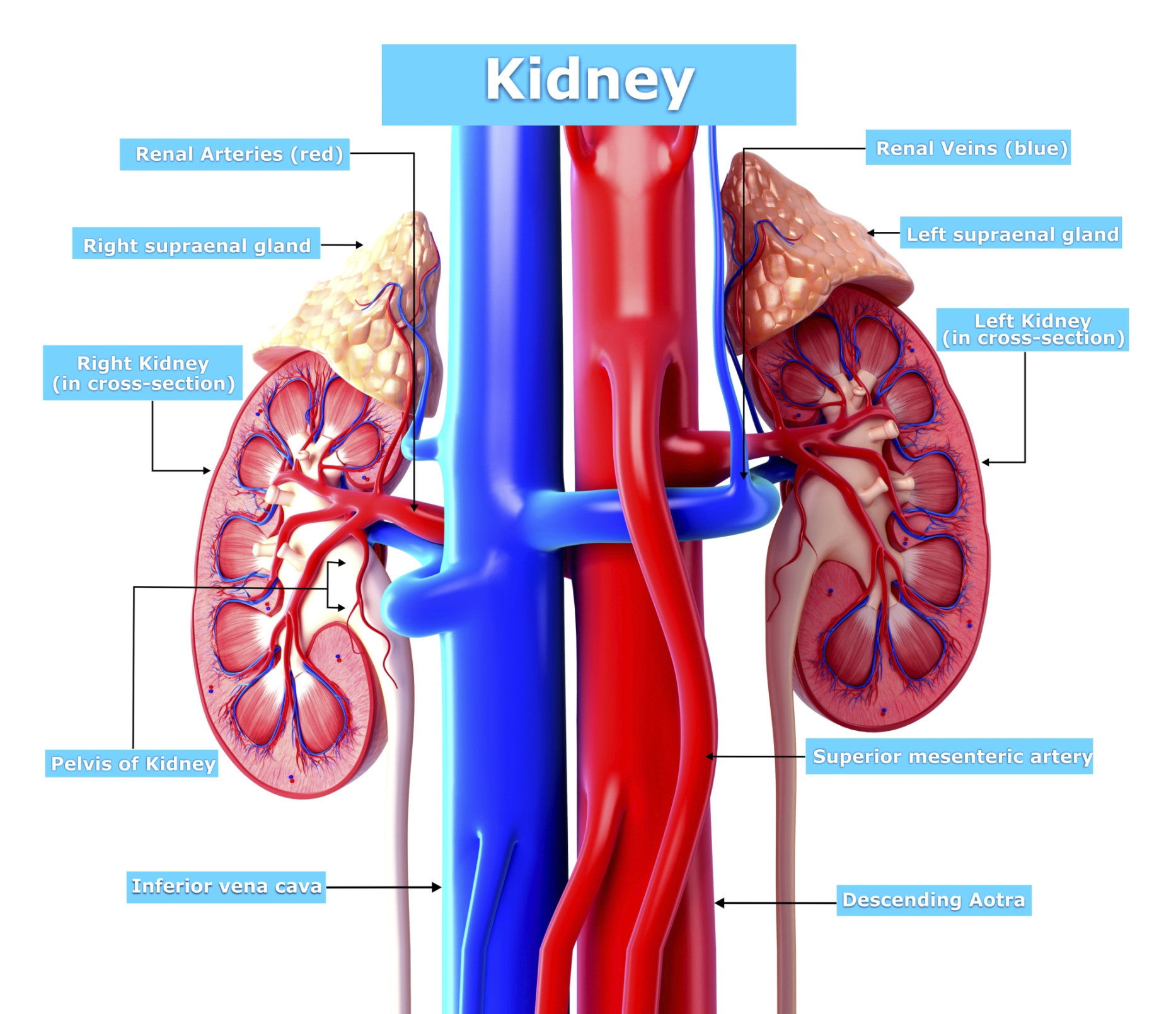
The left kidney is located at about the T12 to L3 vertebrae, whereas the right is lower due to slight displacement by the liver. Upper portions of the kidneys are somewhat protected by the eleventh and twelfth ribs (Figure 25.1.1). Each kidney weighs about 125-175 g in males and 115-155 g in females.

Structure of the kidney medical vector image on VectorStock Medical illustration, Ginjal art
Lobes and Columns. Each kidney contains approximately 6-8 renal lobes, which can be seen without using a microscope. A renal lobe contains thousands of microscopic tubules called nephrons, which are the kidney's functional units. The medullary portion of a renal lobe is called a renal pyramid due to its shape.

Cross Section of Right Kidney Stock Image F031/6574 Science Photo Library
Google Doc Key (TpT) Posted April 5, 2021 in Anatomy, Worksheets by Shannan Muskopf anatomy, kidney, label, learn, nephron, practice, urinary Students practice labeling the urinary system with this drag and drop activity. Three slides have detailed images of the kidneys, ureters, and nephrons.
Kidney Anatomy, Artwork Photograph by Art For Science
Kidney anatomy, unlabeled. Cross section of human kidney showing major structures and location of nephrons. © Alila Medical Media Image size: 21.5 Mpixels (61.5 MB uncompressed) - 5001x4301 pixels (16.6x14.3 in / 42.3x36.4 cm at 300 ppi) Published in: Urinary System Images, Urology Images & Videos
FREE HOMESCHOOLING RESOURCE!!! The Renal System Printable Worksheets
Each kidney weighs about 125-175 g in males and 115-155 g in females. They are about 11-14 cm in length, 6 cm wide, and 4 cm thick, and are directly covered by a fibrous capsule composed of dense, irregular connective tissue that helps to hold their shape and protect them.

Urinary System Kidney Diagram Quizlet
Want to create or adapt books like this? Learn more about how Pressbooks supports open publishing practices.

Kidney Gross Anatomy (Media) Human Bio Media
nephron, functional unit of the kidney, the structure that actually produces urine in the process of removing waste and excess substances from the blood. There are about 1,000,000 nephrons in each human kidney. The most primitive nephrons are found in the kidneys ( pronephros) of primitive fish, amphibian larvae, and embryos of more advanced.

Kidney Diagram Unlabeled Human Body Anatomy
1/3. Synonyms: none. The kidneys are bilateral organs placed retroperitoneally in the upper left and right abdominal quadrants and are part of the urinary system. Their shape resembles a bean, where we can describe the superior and inferior poles, as well as the major convexity pointed laterally, and the minor concavity pointed medially.

45+ Nephron Kidney Anatomy Diagram
Labeling quiz Practice test: Interactive quizzes Sources + Show all Labeled diagram The best way to kick off your revision is with a urinary system diagram which clearly shows all of the structures found within.
Kidney Unlabeled Diagram jkoch526 Flickr
A nephron is the basic structural and functional unit of the kidneys that regulates water and soluble substances in the blood by filtering the blood, reabsorbing what is needed, and excreting the rest as urine. Its function is vital for homeostasis of blood volume, blood pressure, and plasma osmolarity. It is regulated by the neuroendocrine.
